Home

Mispend smal To grader a scan oct Sig til side Generator Dam

Fig. 3.19, [PS-OCT B-Scan through the fovea...]. - High Resolution Imaging  in Microscopy and Ophthalmology - NCBI Bookshelf
Fig. 3.19, [PS-OCT B-Scan through the fovea...]. - High Resolution Imaging in Microscopy and Ophthalmology - NCBI Bookshelf
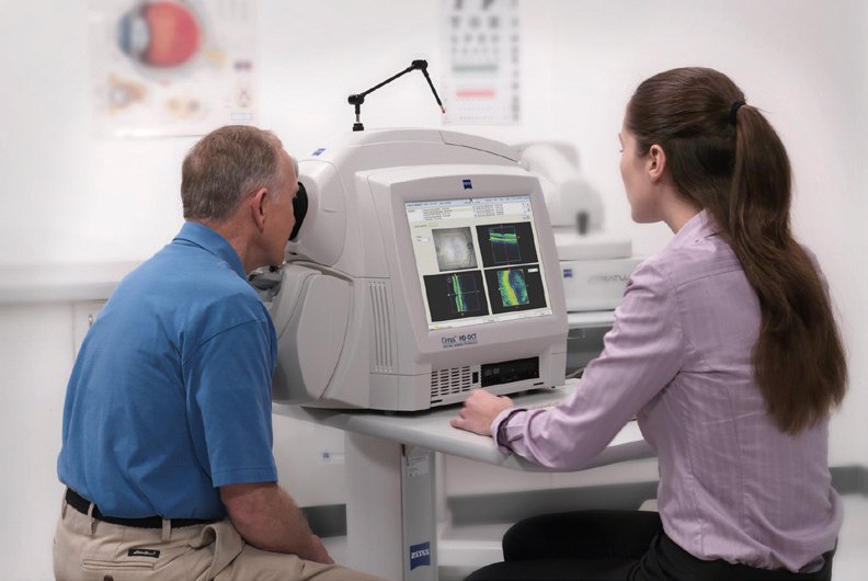
What Is An OCT Scan? | ProVision

A-scan, B-scan and en-face OCT images of the mouse retina. (A)... |  Download Scientific Diagram
A-scan, B-scan and en-face OCT images of the mouse retina. (A)... | Download Scientific Diagram

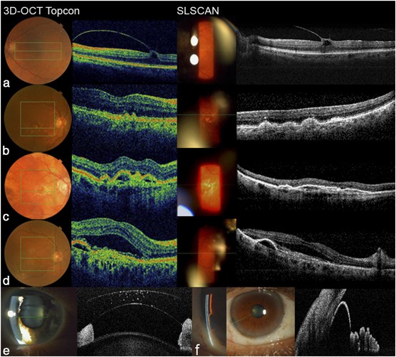
Fourier Domain Optical Coherence Tomography integrated into a slit lamp; a  novel technique combining anterior and posterior segment OCT | Eye
Fourier Domain Optical Coherence Tomography integrated into a slit lamp; a novel technique combining anterior and posterior segment OCT | Eye

Optical Coherence Imaging | Biophotonics Imaging Laboratory | UIUC

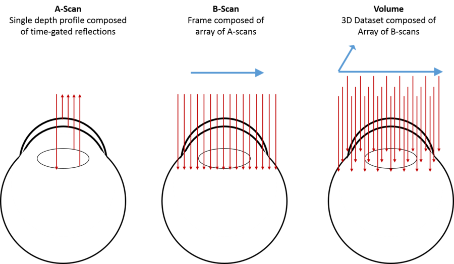
OCT scanning modes. (A) A-line. (B) B-scan. (C) Volumetric OCT. The OCT...  | Download Scientific Diagram
OCT scanning modes. (A) A-line. (B) B-scan. (C) Volumetric OCT. The OCT... | Download Scientific Diagram

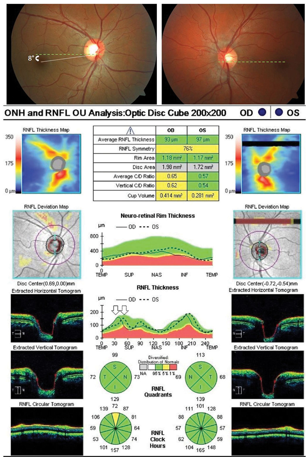
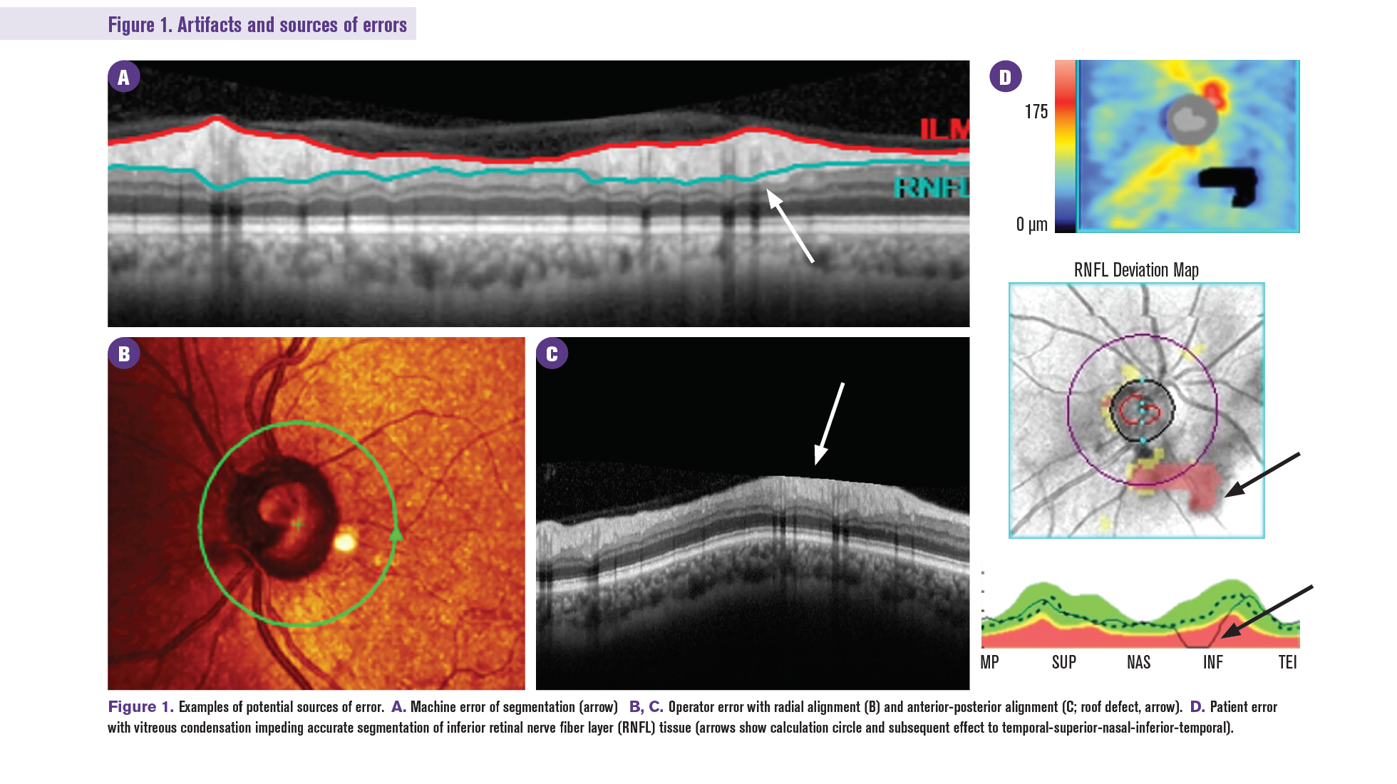
OCT and Glaucoma: Artifact Alert

The Anatomy of an OCT Scan

En-face Flying Spot OCT/Ophthalmoscope | Ento Key

Diagnosing Eye Conditions with Optical Coherence Tomography | FLEI

What is OCT? | Science Lab | Leica Microsystems

PDF] Motion correction in optical coherence tomography volumes on a per A- scan basis using orthogonal scan patterns | Semantic Scholar

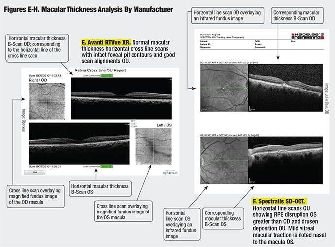
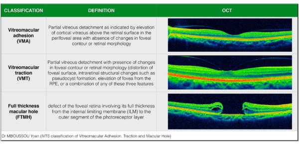
Ophthalmology Management - OCT Terminology — Demystified!

Retinal Physician - Three-Dimensional OCT and OCT Angiography Imaging for  Retinal Diagnosis
Retinal Physician - Three-Dimensional OCT and OCT Angiography Imaging for Retinal Diagnosis

Figure 12.2, OCT B-scan of the retina. Brighter pixels indicate tissue  which reflects more light. The upper portion of the figure shows the  vitreous humor which has very low reflectivity. The small
Figure 12.2, OCT B-scan of the retina. Brighter pixels indicate tissue which reflects more light. The upper portion of the figure shows the vitreous humor which has very low reflectivity. The small

Optical coherence tomography - Wikipedia

5: An OCT A-Scan, B-Scan and C-Scan of a human retina. | Download  Scientific Diagram
5: An OCT A-Scan, B-Scan and C-Scan of a human retina. | Download Scientific Diagram

En Face Optical Coherence Tomography | Ento Key

Anatomy Review - Optical Coherence Tomography Scans

OCT Scanning and scanner coordinate system schematic. Left: 1D... |  Download Scientific Diagram
OCT Scanning and scanner coordinate system schematic. Left: 1D... | Download Scientific Diagram

Classification of healthy and diseased retina using SD-OCT imaging and  Random Forest algorithm | PLOS ONE
Classification of healthy and diseased retina using SD-OCT imaging and Random Forest algorithm | PLOS ONE

12 Ways to Get More Out of Your OCT

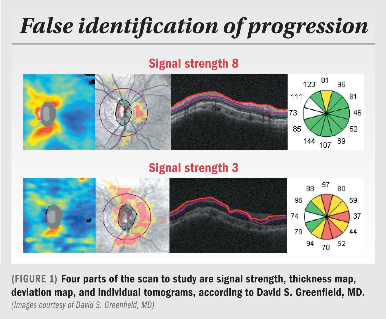
OCT in glaucoma management: Green isn't always clean

Assessing quality of OCT scans impacts clinical-decision process